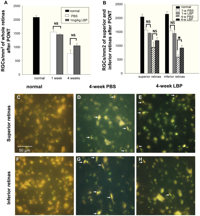
Figure 5. Effects of LBP on RGC survival 1 week and 4 weeks after PONT.
The RGCs were labeled with FG. (A) LBP did not increase the survival of RGCs either 1 week or 4 weeks after the PONT when the densities of surviving RGCs were produced from the whole retinas (NS: not significant). (B) When the retinas were divided into the superior and inferior halves, LBP did not delay the degeneration of RGCs 1 week after PONT. However, it reduced the degeneration of RGCs in the inferior retina (*P = 0.027) but not in the superior retina 4 weeks after the PONT. (F – H) The photographs of RGCs labeled by FG in both the superior and inferior retinas are about 1.5 mm away from the optic disc. In the superior retinas, the densities of RGCs were similar between the PBS and LBP groups. In the inferior retinas, the density of RGCs in the LBP group was higher than that in the PBS group. Microglia (white arrows) were easily distinguished from RGCs and not counted. (n = 7 and 4 in PBS and LBP groups 1 week after PONT. n = 9 and 10 in PBS and LBP groups 4 weeks after PONT.).

