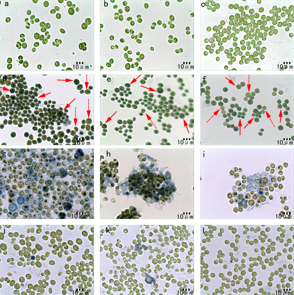
Figure 7.
Microscopic pictures of microalgal cells: controlled cells: a) Chlorococcum nivale; b) Chlorococcum ellipsoideum; c) Scenedesmus sp.; cells heated at 121 and incubated in 1% Evans’ blue solution for 3 h: d) Chlorococcum nivale; e) Chlorococcum ellipsoideum; f) Scenedesmus sp.; cells flocculated by adjusting pH value of growth medium to 0.5 with nitric acid and incubated in 1% Evans’ blue solution for 3 h: g) Chlorococcum nivale; h) Chlorococcum ellipsoideum; i) Scenedesmus sp.; cells flocculated by adjusting pH value of growth medium to 3.5 with nitric acid and incubated in 1% Evans’ blue solution for 3 h: j) Chlorococcum nivale; k) Chlorococcum ellipsoideum; l) Scenedesmus sp.

