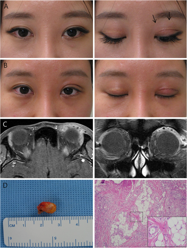
Figure 2.

Case 2. A) Preoperatively, this patient had small nodoular masses with mild lagophtlmos. B) Two months postoperatively, no mass remained and mild lagophthalmos was improved. C) Coronal and axial enhanced MRI views showing diffuse eyelid enhancing. D) The largest excised masses (9×7 mm) of Case 2. Microscopically, the specimen contained chronic inflammation with foreign body lipogranuloma (H&E stain, ×100, ×400 with magnification).
