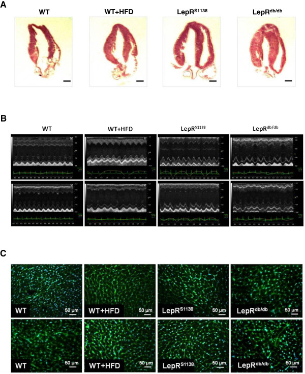
Figure 1.
Cardiac phenotype of lean and obese WT, WT + HFD, LepRS1138 and LepRdb/db mice. (A) Representative H&E-stained longitudinal sections through hearts of 7 months-old mice are shown. Magnification, ×10. (B) Representative M-mode echocardiographic recordings. (C) Representative images of wheat germ agglutinin (WGA)-stained myocardial cross sections. The mean cardiomyocyte cross-sectional areas are given in Table 1.

