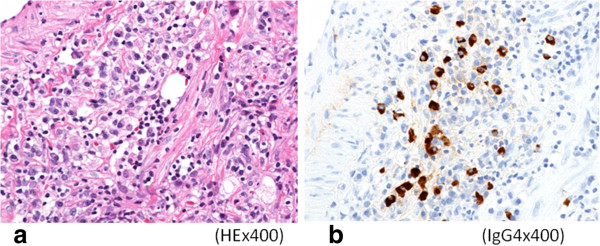
Figure 4.

Microscopic examination. a) Histopathological examination revealed pronounced inflammatory cell infiltration consisting largely of plasma cells, macrophages and lymphocytes on a background comprised of fibrous interstices with fibrosis and fibroblast proliferation. b) Immunohistochemical examination revealed for IgG4 showed a high IgG4/IgG ratio (×400).
