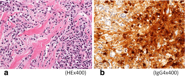
Figure 7.

Microscopic examination. a) Pathological examination of the lung revealed marked lymphocytic infiltration in the vicinity of alveolar epithelium free of atypia and marked interstitial connective tissue proliferation with hyaline degeneration. b) Immunohistochemical examination revealed for IgG4 showed a high IgG4/IgG ratio, exceeding 60%.
