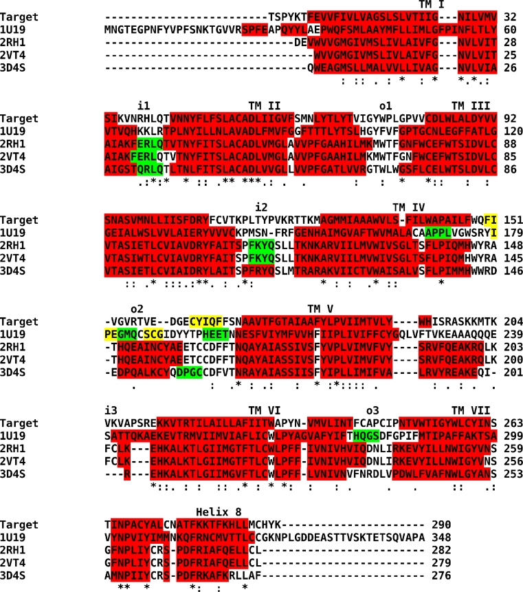
Fig. 1.
Alignments of templates to target structure. Alignment of templates for homology modeling labeled by their PDB entry code to the target sequence of the human M2 muscarinic acetylcholine receptor. Stars denote conserved and dots consensual residues. Colors denote secondary structure: red—helix; white—coil; yellow—strand; green—turn. Secondary structure of the target was predicted by PsiPred (http://bioinf.cs.ucl.ac.uk/psipred/) and secondary structures of templates were taken from respective crystal structures. For orientation transmembrane (TM) helices, inner (i) and outer (o) loops are indicated

