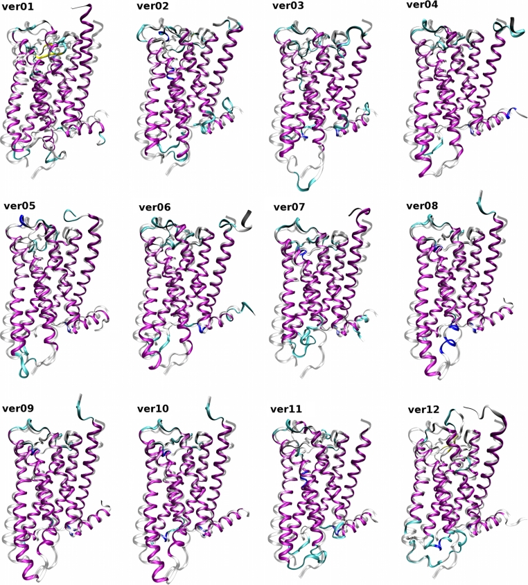
Fig. 2.
Twelve homology models of the M2 receptor superposed on the 3UON crystal structure. Homology models (color) of the M2 receptor based on the templates listed in Table 1 are superposed using MUSTANG [27] implemented in YASARA on the crystal structure 3UON (gray). Orientation: extracellular site up, TM VI and TM VII front. Colors: purple—α-helix; yellow—β-sheet; cyan—turn; white—coil. RMSDs of the models to the target structure (3UON) are listed in Table 4

