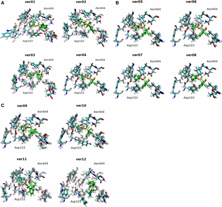
Fig. 5.
The best QNB docking poses of 12 homology models superposed on the 3UON crystal structure. View from the extracellular site, TM II down, TM VI and TM VII up, of the best docking poses, according to the binding energy estimates (Fig. 4), of QNB (green carbons) and residues of the orthosteric binding site (color) superposed on the crystal structure of 3UON (gray). Colors: Cyan—carbon; red—oxygen; blue—nitrogen; white—hydrogen; yellow—hydrogen bonds. The residue labels correspond to the M2 sequence. The calculated RMSDs of QNB and residues of the orthosteric binding site of the models to the target structure (3UON) are shown in Table 5

