Fig. 2.

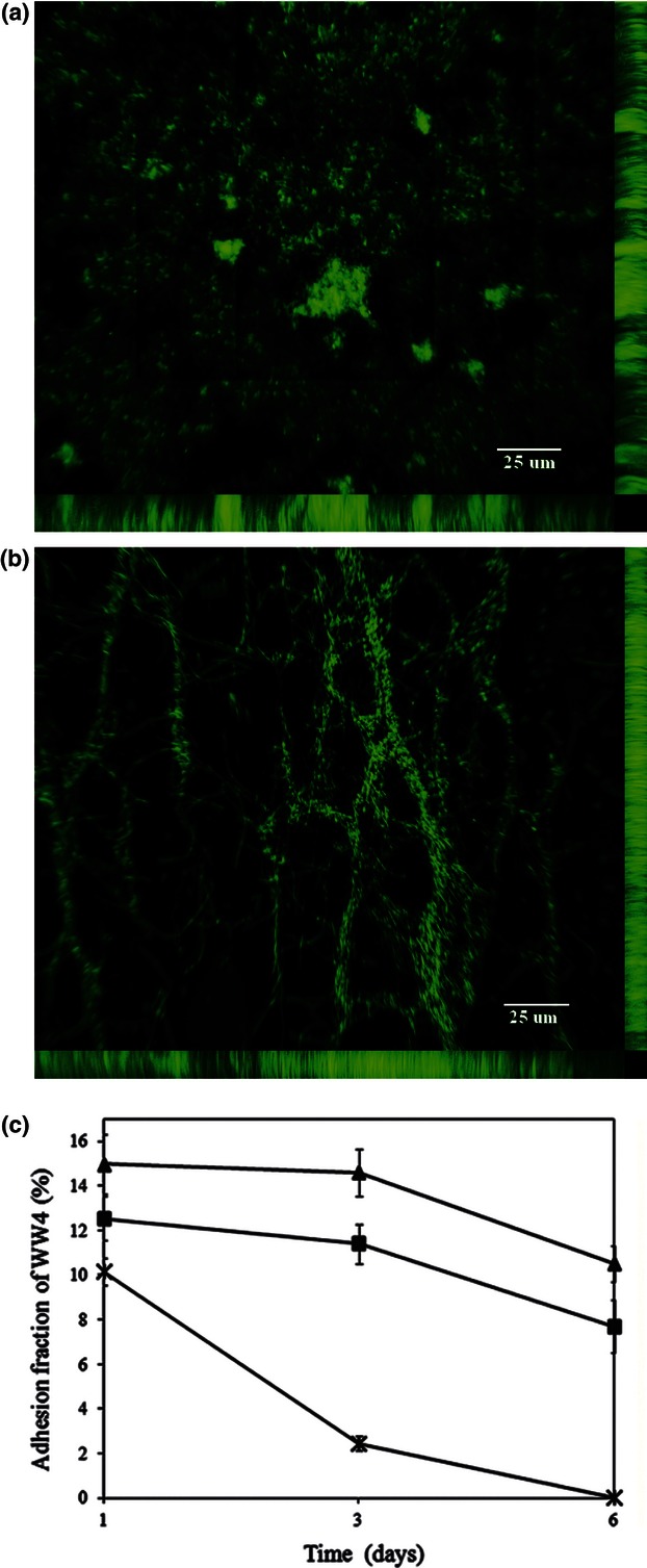
The biofilm of Serratia marcescens WW4 and P. aeruginosa WW5 in chambers, and the adhesion fraction of S. marcescens WW4 in pure or co-cultures. The hill-like biofilm structure of P. aeruginosa WW5 (a) and the filamentous biofilm structure of S. marcescens WW4 (b) were observed in BM medium in the biofilm-forming chambers. The pictures were obtained from 1-day DAPI-stained cultures, and the bars represent 25 μm. (c) The biofilm of S. marcescens WW4 mixing in the absence (×) or presence of equal (▲) or tenfold (▪) P. aeruginosa WW5 was incubated in phosphate-limited chambers. The adhesion fraction of S. marcescens WW4 was photographed at 1, 3 and 6 days using a fluorescence microscope, and the images were analysed using ImageJ software. All data show the means and standard errors of the adhesion proportion (%) of S. marcescens WW4 biofilm.
