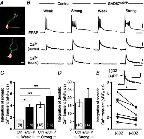
Figure 5. Ca2+ transients in the PC soma induced by activation of weak CFs are larger in GAD67+/GFP mice than in control mice.

A, representative PC images of a control (upper) and a GAD67+/GFP (lower) mouse. Areas indicated by red and green dotted lines represent ROIs for somatic and dendritic Ca2+ transients, respectively. Scale bars, 20 μm. B, EPSPs and Ca2+ transients recorded in the soma and dendrite of multiply innervated PCs in response to stimulation of a weak or a strong CF in a control and a GAD67+/GFP mouse. Scale bars, 20 mV and 10 ms for EPSPs, 2% (for soma) or 10% (for dendrite) and 1 s for Ca2+ transients. C, D, average magnitudes of Ca2+ transients from the PC soma (C) and dendrites (D) induced by stimulating a weak (CF-multi-W) or a strong (CF-multi-S) CF. Ca2+ transients for 1.5 s from the onset were integrated. For the data in C, statistical analysis was performed using the Kruskal–Wallis test with the Steel-Dwass multiple comparison post hoc test. *P < 0.05, **P < 0.01, ***P < 0.001. E, traces show somatic Ca2+ transients elicited by activation of weak CF in GAD67+/GFP PCs before (–) and after (+) bath application of diazepam (DZ, 1 μm). Data from each cell are plotted in the lower graph. *P < 0.05. Scale bars, 2% and 1 s. Modified with permission from Nakayama et al. (2012).
