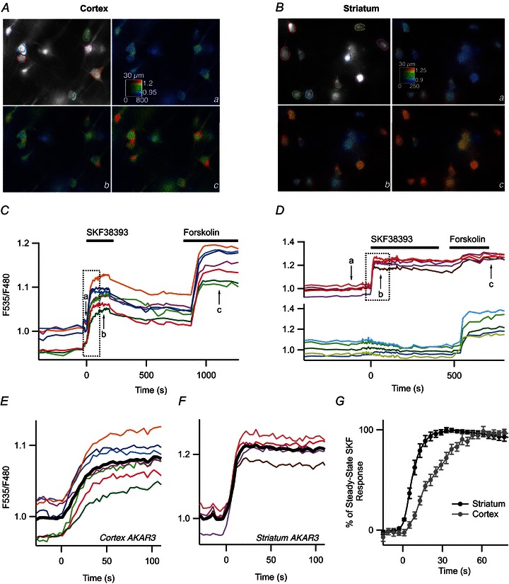
Figure 5. The onset of the PKA response is faster in the striatum than in the cortex.

SKF38393 (1 μm) was applied with a fast perfusion system. Pyramidal cortical neurones (A) and MSNs in the dorsal striatum (B) were transduced for AKAR3 expression and imaged by wide-field microscopy. Images show the raw fluorescence at 535 nm (left, in grey scale) and the ratio (in pseudocolour), indicating levels of PKA-dependent phosphorylation in control condition (a), during the responses to 1 μm SKF38393 (b) and 13 μm forskolin (c). C and D, time-course of the F535/F480 emission ratio measured in the regions indicated by the colour contour on the grey-scale image in the cortex (A) and striatum (B), respectively. E and F, expansion indicated by the dotted rectangle in of C and D, respectively, showing the response onset. The black trace represents the average. G, average similar experiments in the cortex (n= 8) and striatum (n= 10). Error bars represent SEM.
