
Figure 7. Brief (10 s) applications of SKF38393 produced stronger and longer-lasting PKA responses in the striatum than in the cortex.
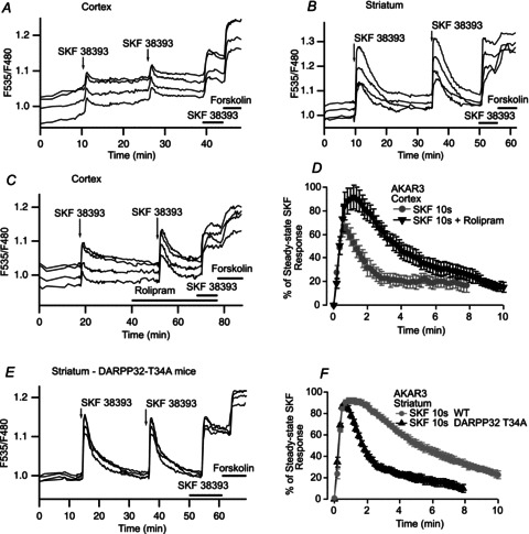
PKA activation was measured with AKAR3 during the application of a 10 s pulse of 1 μm SKF38393 in the cortex (A) and striatum (B). Each trace on the graphs indicates the AKAR3 emission ratio of an individual neurone in wide-field imaging. Responses were reproducible, with no significant difference in amplitude or kinetics. At the end of each experiment, SKF38393 and forskolin (13 μm) were added to the bath for the times indicated by the horizontal bars, and the response to the sustained presence of SKF38393 was used for normalisation. C, PKA response to a brief (10 s) application of SKF38393 (1 μm) alone and in the presence of the PDE4 inhibitor rolipram (100 nm), in the cortex: rolipram strongly increased the amplitude and prolonged the duration of the PKA response. D, mean responses in the cortex to brief SKF38393 stimulation alone (n= 6) or in the presence of rolipram (n= 4); bars indicate the SEM. E, PKA response to a brief (10 s) application of SKF38393 (1 μm) in the striatum of DARPP-32 T34A mice. F, mean responses to brief SKF38393 stimulation, in the striatum of wild-type (n= 7) and DARPP-32 T34A mice (n= 7). The higher level of phosphatase 1 activity in DARPP-32 T34A mice had no effect on the amplitude of the PKA response, but strongly reduced the duration of the PKA response.