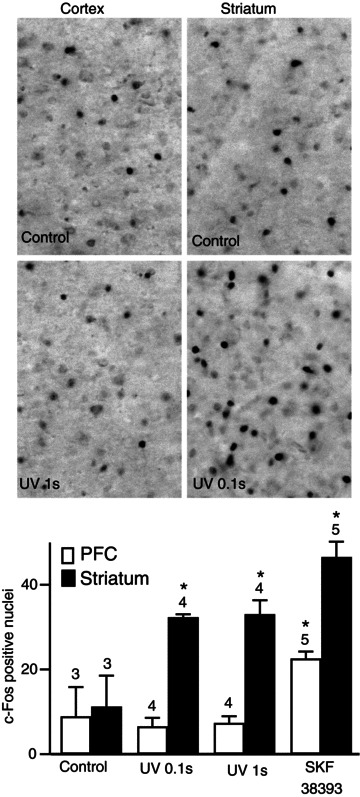
Figure 9. A sub-second dopaminergic stimulation was sufficient to induce c-Fos expression in the striatum, but not in the cortex.

PFC, prefrontal cortex. Brain slices were stimulated with dopamine released by the local flash photolysis of NPEC-dopamine (5 μm, 0.1 or 1 s) or bath application of SKF38393 (1 μm, 5 min). The brain slice was fixed 90 min after the stimulus and c-Fos-immunoreactive nuclei were quantified in both structures. Grey-scale images show brain slices stained for c-Fos in the conditions indicated in the panel. Positive nuclei were counted and the counts were plotted on the histogram: each bar indicates the mean number of c-Fos-positive nuclei in the corresponding conditions and error bars indicate the SEM. Significant responses were obtained for all uncaging durations for the striatum, whereas the cortex responded only to bath-applied SKF38393. Unpaired two-tailed t tests were carried out for comparisons with control conditions, and differences were considered significant when P < 0.05, indicated by an asterisk on the graph.
