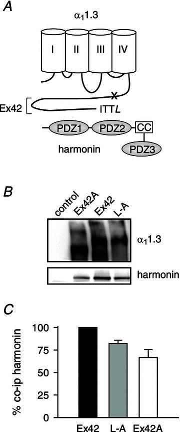
Figure 3. Harmonin binding is reduced by disruption of the PDZ-binding site in the α11.3 dCT.

A, schematic diagram of harmonin and α11.3. The long C-terminal domain encoded by exon 42 (Ex42) includes the PDZ-binding motif (ITTL) that interacts with the second of three PDZ domains of harmonin. The coiled-coil domain (CC) that is deleted in the dfcr harmonin mutant is indicated. The ‘x’ marks approximate location of truncation of the dCT due to inclusion of exon 42A. B, coimmunoprecipitation of harmonin with Cav1.3 channels in transfected HEK293T cells. Cells were transfected with harmonin alone (control) or cotransfected with Cav1.3 subunits including α11.3 with full-length (Ex42) or truncated (Ex42A) dCT or α11.3 with terminal leucine substituted with alanine (L-A). Western blotting detected α11.3 (upper panel) and coimmunoprecipitated harmonin (lower panel). C, quantification reflecting percentage change in harmonin coimmunoprecipitated with α11.3Ex42A and α11.3L-A compared to that for α11.3Ex42 (%co-ip harmonin). See Methods for details.
