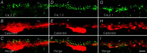
Figure 3. Immunohistochemical localization of N-, L-, and P/Q-type Ca channels in horizontal cells.

A, fluorescence micrograph of labelling by antibodies to N-type Ca channel α1B subunits (Cav2.2, in green). B, fluorescence micrograph of labelling of calbindin, a cell marker for horizontal cells in the outer plexiform layer, on the same section (in red). C, the merged image of these two images. D. labelling of L-type Ca channel α1C subunits (Cav1.2, in green). E, labelling of calbindin (in red). F, the merge of these two images. G, labelling of P/Q-type Ca channel α1A subunits (Cav2.1, in green). H, the same section labelled for calbindin. I, the merge of these two images. The merged images indicate co-localization of calbindin and Ca channel immunolabelling. Each image is a projection of three optical sections. Asterisks denote horizontal cell bodies, and the arrows point to the invaginating tips of horizontal cell processes. Scale bar, 5 μm.
