
Figure 2. Sanshool suppresses mechanosensitivity of cutaneous somatosensory fibres.
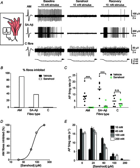
A, example recordings demonstrate the inhibition of mechanically evoked action potentials in SA-Aβ and AM fibres in the presence of sanshool. B, percentage of nerve fibres that exhibit mechanical inhibition in sanshool. P < 0.001 (χ2 test). n= 8–10. C, number of action potentials fired at baseline and after 3 min in sanshool for each fibre tested. Solid bar represents the mean. ***P < 0.001, (two-way ANOVA). n= 8–10. D, sanshool concentration–inhibition curve for AM fibres using a 100 mN force stimulus. Data were fitted by a non-linear four-parameter regression model. E, dose-dependent inhibition of AM fibres by sanshool in response to increasing mechanical force. n= 9–14. AM, Aδ mechanoreceptor; AP, action potential; C, C fibre; n.s., not significant; SA, slowly adapting.