
Figure 3. mRNA expression of Na pump α subunits in the cerebellum of juvenile mouse (postnatal day 40).
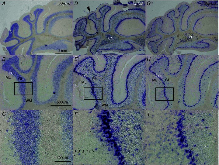
Alternate sections were examined by in situ hybridization using different probes. A–C, Atp1a1 mRNA expression. D–F, Atp1a2 mRNA expression. Atp1a2 mRNA expression in the pia matter (arrowhead). G–I, Atp1a3 mRNA expression. Boxes in B, E and H show areas that are pictured beneath at higher magnifications (C, F and I, respectively). In each panel, the top is the dorsal and the right is the medial side. CN, cerebellar nuclei; ML, molecular layer; GL, granular layer; PCL, Purkinje cell layer; WM, white matter.