
Figure 4. Expression of Na pump α3 subunits, VGAT, GAD65/67, VGLUT1 and VGLUT2 in the cerebellum of young wild-type mice.
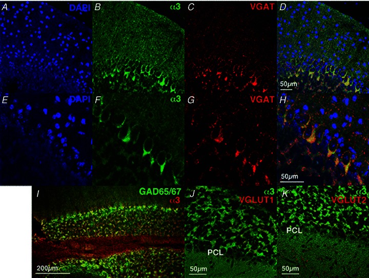
Immunofluorescence using antibodies to α3 (green, B and F) and VGAT (red, C and G). Nuclei are stained with DAPI (blue, A and E). E–H, higher magnifications of A–D. Merged figures of α3 and VGAT are shown in D and H. A merged figure of immunofluorescence using antibodies to α3 (red) and GAD65/67 (green) is shown in I. Merged figures using antibodies to α3 (green) and VGLUT1 (red, J) or VGLUT2 (red, K) are shown. A–I, P26 mice; J and K, P39 mice.