
Figure 7. Comparison of parallel fibre (PF)- and climbing fibre (CF)-mediated excitatory neurotransmission onto PCs between WT and Atp1a3+/−.
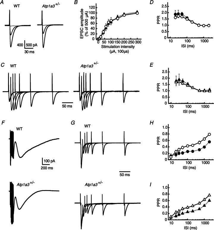
A, representative averaged traces of PF-mediated EPSCs recorded from a single PC of WT and Atp1a3+/−. The EPSCs were evoked by electrical stimulation with a series of different intensity (from 10 to 500 μA for 100 μs), and superimposed for stimulations at 10 (pale grey lines), 60 (dark grey lines) and 500 μA (black lines). Each trace is derived from averaging the EPSCs of several successive traces recorded every 15 s. Stimulation artifacts were truncated for clarity. B, relationship between the amplitude of PF-PC EPSCs and stimulation intensity for WT (open circles, n= 14) and Atp1a3+/− (filled triangles, n= 13). Data are mean ± SEM and are relative to the amplitude examined at a stimulation intensity of 500 μA. C, representative averaged traces of PF-PC EPSCs examined by the paired-pulse protocol in PCs of WT and Atp1a3+/−. The traces were normalized relative to the peaks of the first EPSC. D and E, relationship between the PPR and ISI for PF-PC EPSCs examined in WT (D, circles) and Atp1a3+/− (E, triangles). The magnitudes of PPR of the EPSC were not significantly different between WT and Atp1a3+/− at any interval tested in the absence (open symbols) and presence (filled symbols) of 2 mmγ-DGG and 50 μm cyclothiazide. F, representative averaged traces of mGluR1/TRPC1-mediated inward currents examined in PCs of WT and Atp1a3+/−. The currents were evoked by trains of PF stimuli (100 Hz for 100 ms) in the presence of 20 μm CNQX and 100 μm picrotoxin. G, representative averaged traces of CF-EPSCs examined by the paired-pulse protocol in PCs of WT and Atp1a3+/−. The traces were normalized relative to the peaks of the first EPSC. H and I, relationship between the PPR and ISI for CF-PC EPSCs examined in WT (H, circles) and Atp1a3+/− (I, triangles). The PPR of CF-EPSCs was not different between WT and Atp1a3+/−, when they were compared in the absence (open symbols) and presence (filled symbols) of γ-DGG and cyclothiazide.