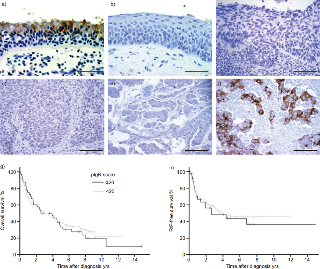
FIGURE 1.
Polymeric immunoglobulin receptor (pIgR) expression in normal bronchial epithelium, pre-invasive lung lesions and lung cancers evaluated by immunohistochemistry (IHC). a) IHC on normal lung tissues revealed a strong staining for pIgR in mucous and serous cells in surface and glandular epithelium, a weak staining in ciliated cells, and an expected absence of staining in alveolar cells. b, c) There was no pIgR expression in pre-invasive lung lesions: b) squamous metaplasia; c) severe dysplasia. pIgR expression was also absent in d) 90% of squamous cell carcinomas and e, f) 52% of adenocarcinomas (ADCs): e) ADC with absence of pIgR staining and f) ADC with strong pIgR staining. Scale bars=20 µm. Kaplan–Meier analysis of g) overall survival (p=0.66) and h) recurrence (R)- or progression (P)-free survival (p=0.67) did not show a survival difference between lung cancer patients with or without pIgR expression.

