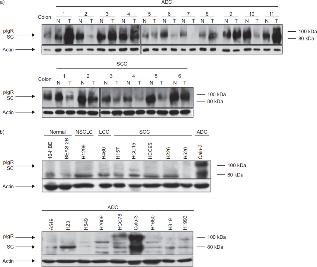
FIGURE 2.
Polymeric immunoglobulin receptor (pIgR) expression in lung tissues and cell lines evaluated by immunoblotting. Whole tissue or cell lysates were resolved by sodium dodecylsulfate–polyacrylamide gel electrophoresis and blots were incubated with anti-pIgR antibody. pIgR expression levels were normalised to actin. a) Lung cancer tissues (T) were compared to normal lung tissues (N) from the same patients. The results show downregulation of pIgR expression in six out of six squamous cell carcinomas (SCCs) and in five out of 11 adenocarcinomas (ADCs), while pIgR was overexpressed in four out of 11 ADCs and no expression difference was noted in two out of 11 ADCs. b) pIgR expression was low in two out of two transformed normal bronchial epithelial cell lines, one out of one undifferentiated nonsmall cell lung cancer (NSCLC) cell line, one out of one large cell carcinoma (LCC) cell line, five out of five SCC cell lines and seven out of nine ADC cell lines. pIgR expression was moderate to strong in two out of nine ADC cell lines. SC: secretory component.

