FIGURE 3.
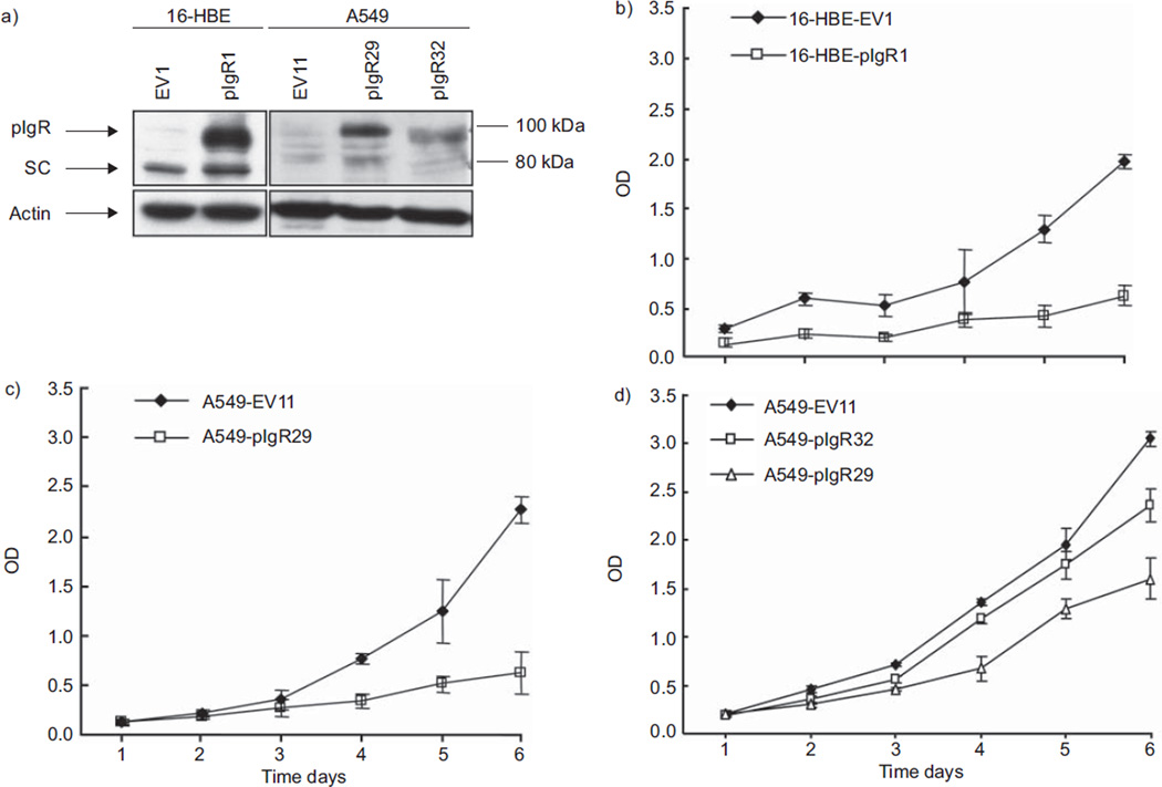
Effects of polymeric immunoglobulin receptor (pIgR) overexpression on 16-HBE and A549 proliferation. a) Whole cell lysates were resolved by sodium dodecylsulfate–polyacrylamide gel electrophoresis and blots were incubated with anti-pIgR antibody. pIgR expression levels were normalised to actin. Higher expression of pIgR was confirmed by immunoblotting in immortalised normal bronchial epithelial cell line 16-HBE and lung adenocarcinoma cell line A549 stably transfected with pIgR pcDNA3.1(−) (16-HBE-pIgR1, A549-pIgR29 and A549-pIgR32) as compared with those transfected with the empty vector (16-HBE-EV1 and A549-EV11). pIgR expression was higher in A549-pIgR29 (clone with the strongest pIgR expression) than in A549-pIgR32 (clone with moderate pIgR expression). SC: secretory component. b) Inhibition of cell growth was observed by WST-1 assay in 16-HBE-pIgR1 grown for 6 days as compared with 16-HBE-EV1. Optical density (OD) in the y-axis reflects the proportion of metabolically active cells. c) Inhibition of cell growth was observed by WST-1 assay in A549-pIgR29 grown for 6 days as compared with A549-EV11. d) Dose-dependent effect on cell growth inhibition in A549 cells. Inhibition of cell growth was less pronounced in A549-pIgR32 (clone with moderate expression of pIgR) than in A549-pIgR29 (clone with higher expression of pIgR). Error bars represent standard deviation (n=5). All the graphs represent one of three independent experiments with similar results.

