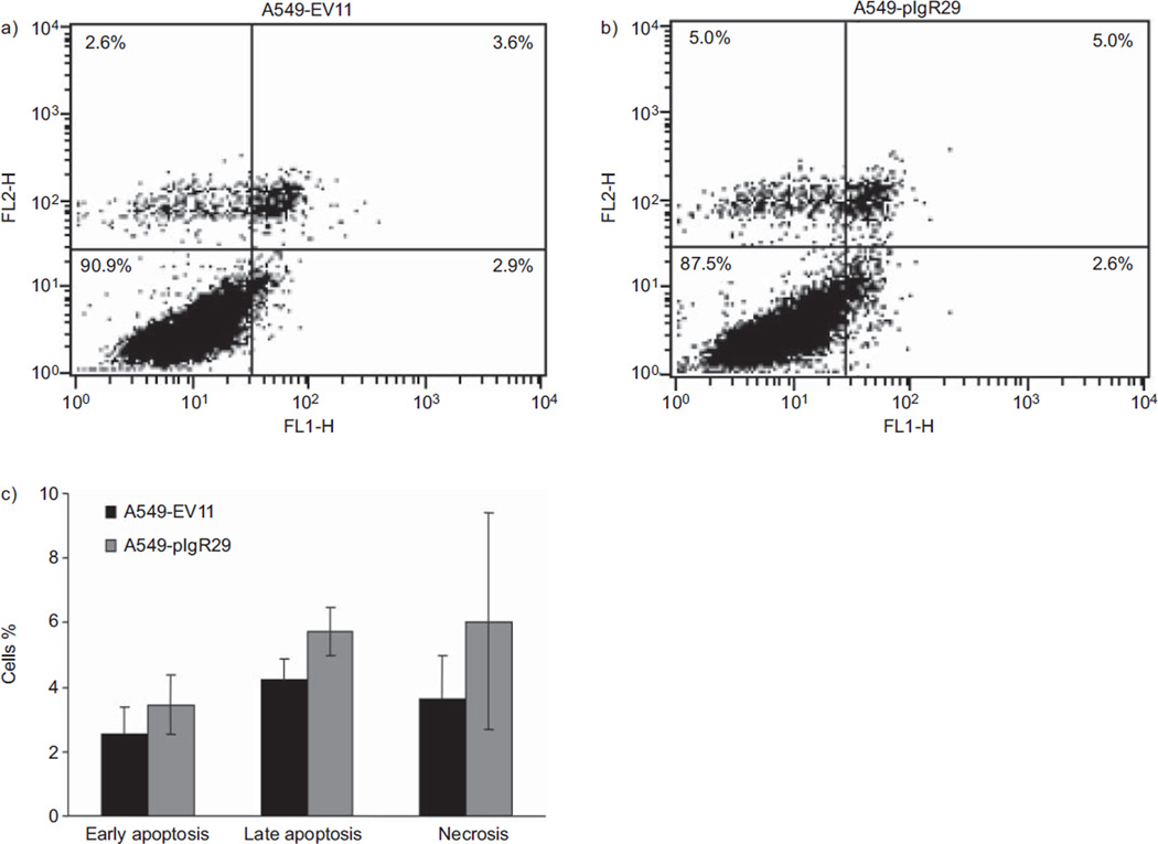
FIGURE 5.
Effects of polymeric immunoglobulin receptor (pIgR) overexpression on A549 apoptosis evaluated by annexin V apoptosis assay. Stably transfected cells were stained with annexin V and propidium iodide (PI), and the staining was quantified by fluorescence-activated cell sorting analysis. Quadrant dot blot analysis of a) A549-EV11 and b) A549-pIgR29 cells stained for apoptosis using an annexin V detection of phosphatidyl serine expression along the x-axis, counterstained with PI to detect late apoptotic or necrotic cells along the y-axis. In the lower left quadrant, viable nonapoptotic cells are found, and in the lower right quadrant, apoptotic cells binding annexin V. Cells in the upper right are late apoptotic cells binding annexin V and taking up PI, and in the upper left are necrotic cells taking up PI but not binding annexin V. These figures are representative of three independent experiments. c) The spontaneous level of early apoptosis, late apoptosis and necrosis had a tendency to be higher in cells overexpressing pIgR (A549-pIgR29), but the difference was not statistically significant. Average of three independent experiments is shown. Error bars represent standard deviation (n=3).

