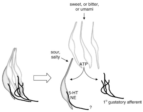
Fig. 4.
Schematic diagram of hypothesized signal processing in mammalian taste buds. At left is shown a postulated taste bud processing unit, consisting of a cluster of Receptor (Type II) Cells, a Presynaptic (Type III) Cell, and their nerve innervation. For simplicity, only 1 Presynaptic Cell is illustrated. Type I ensheathing glial-like cells that may surround the cluster and limit ATP diffusion have also been omitted for clarity. At the right is shown the same cluster, but expanded to illustrate cell-to-cell communication mediated by taste-evoked ATP. Taste excitation by sweet, bitter or umami compounds stimulate Receptor cells to secrete ATP and activate 1° gustatory afferent fibers. Taste-evoked ATP also excites Presynaptic Cells which form synapses with as-yet unidentified fibers (shown by “?”). Sour and salty stimuli act directly on Presynaptic Cells [27]. Presynaptic Cells release serotonin and noradrenalin, perhaps as neurocrine transmitters at their synapses, or as paracrine transmitters acting within the confines of the taste bud, or both.

