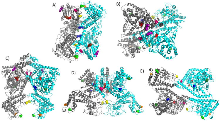
Figure 1.
SecA dimer crystal structures with monocysteine residues colored. The color assignments for the residues are as follows: 59-dark green, 340-yellow, 402-blue, 427-hot pink, 458-brown, 470-purple, 506-red, 696-green, and 734-orange. E. coli amino acid residues are given and homologous residues in other species are depicted based on sequence alignment. For each dimer, the individual protomers are shown in grey or cyan. (A) Hunt et al. B. subtilis structure (PDB ID: 1M6N) 21, (B) Zimmer et al. B. subtilis structure (PDB ID: 2IBM) 25, (C) Vassylyev et al. T. thermophilus structure (PDB ID: 2IPC) 24, (D) Papanikolau et al. E. coli structure (PDB ID: 2FSF)22, and (E) Sharma et al. M. tuberculosis structure (PDB ID: 1NL3) 23.

