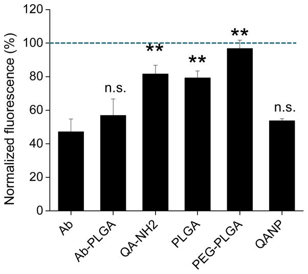
Fig. 2.
Binding-inhibition assay. Fluorescence of HL-60 bound to HUVEC, which were pre-treated with NPs (0.1 mg/mL), QA-NH2 (20 mM), or E-selectin Ab (5 μg/mL). 0.1 mg/mL QANPs was equivalent to 2 μM of QA. The fluorescence obtained with each treatment is normalized to the fluorescence of HL-60 bound to serum-starved medium (SSM)-treated HUVEC. Therefore, NPs or ligands interfering with the HL-60-HUVEC binding results in fluorescence lower than 100% (SSM-treated HUVEC, dotted line). Data are expressed as average ± standard deviation of 3–6 independently obtained results per treatment. **: p<0.01 vs. Ab; n.s.: not significant, p>0.05 vs. Ab.

