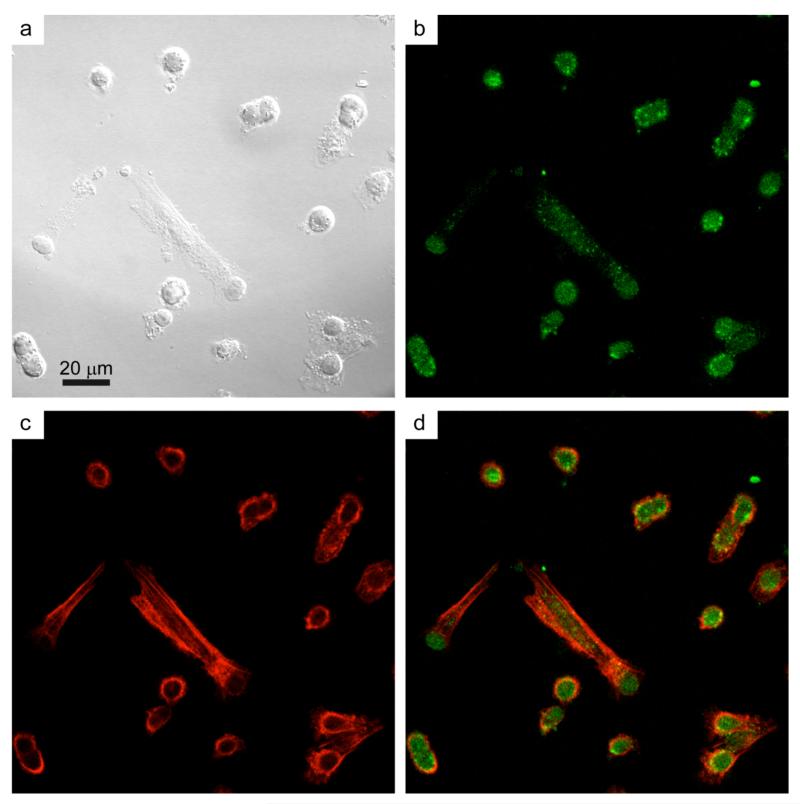
Figure 6.
Uptake of EGF-functionalized Y2O3:Tb3+ nanophosphors by VE11 mouse melanoma cells. VE11 cells were incubated for 4 hours with EGF-functionalized nanoparticles and then fixed and stained with FITC-phalloidin to reveal the actin cytoskeleton. (a) Differential interference contrast (DIC) image of VE11 mouse melanoma cells after 4 hours of incubation with EGF-functionalized Y2O3:Tb3+ nanophosphors. (b) Confocal image of the nanophosphors emission and (c) the actin cytoskeleton. (d) Merged image of panels (b) and (c).

