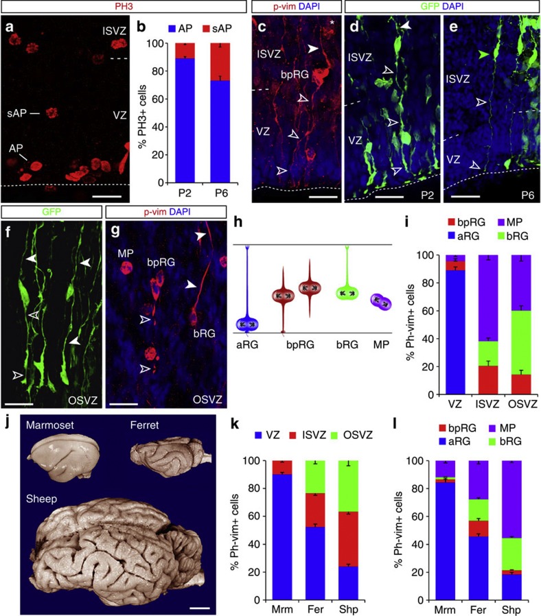
Figure 4. Distribution of mitotic cells in the neocortex of gyrencephalic species shows a high abundance of bipolar radial glia.
(a,c–g) Fluorescence micrographs of ferret neocortex sections (postnatal day 2, P2 (c,d,f,g) and P6 (e) immunostained as indicated (phosphorylated histone H3=PH3; phosphorylated-vimentin=p-vim)), showing mitotic apical (AP) and subapical progenitors (SAP) within the VZ and ISVZ (a,c–e) and OSVZ (f,g). Progenitor cells in ISVZ and OSVZ displaying the typical morphology of bpRGs with a basal process (solid arrowheads) and a long apical process (open arrowheads) were revealed by Rv:gfp (d,f) or Adeno-gfp injection (e) or p-vim (c,g). Note also bRGs with only a basal process and multipolar progenitors (MP) without longer processes. Schematic drawings in (h) depict the defining characteristics of the different progenitor cell types analysed in the species indicated in (j); external view of the adult brains at scale. Histograms in (b,i,k,l) depict AP and bpRG, MP and bRG mitoses in the ferret VZ at P2 and P6 (b), n=982, 250, respectively (three animals each), proportions of the cell types indicated in (h) defined by p-vim in P2 ferret neocortex (i), n=623 cells in VZ, 290 ISVZ, 277 OSVZ (three animals), and in all germinative layers (l), the contribution of which is shown in (k), in the marmoset (Mrm), ferret (Fer) and sheep (Shp) at equivalent stages of cortical development (E85, P2, E65–67, respectively; marmoset, n=1,743 cells; ferret, 1,190 cells; sheep, 2,435 cells; three subjects of each species). Data are mean±s.e.m. Scale bars, 20 μm (a,c,d,f,g); 40 cm (e); 1 cm (j).

