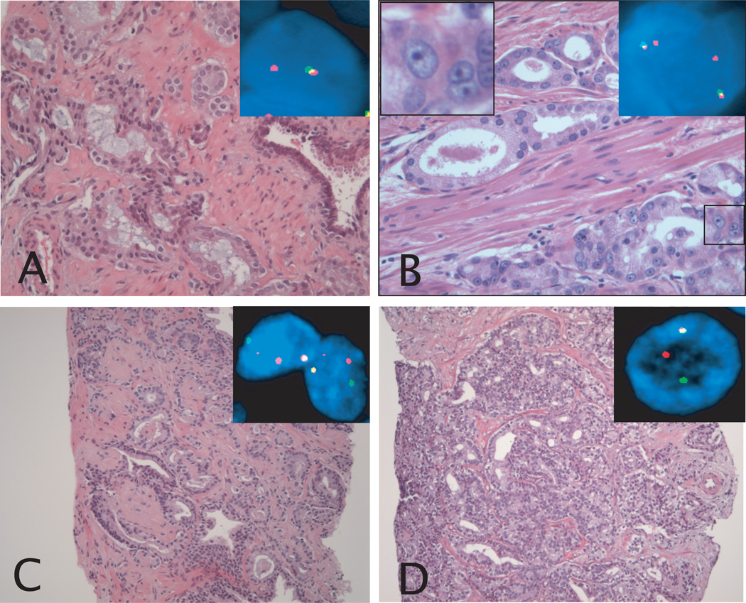
Figure 1.
Morphological features associated with a positive TMPRSS2-ERG fusion status in prostate cancer. A: Prostate cancer Gleason pattern 3 showing blue-tinged mucin. Inset picture shows FISH image of representative nucleus. One yellow and one red signal are present, demonstrating the presence of TMPRSS2-ERG fusion through deletion. B: Prostate cancer Gleason pattern 3 showing macronucleoli. The inset picture in the upper left corner shows the macronucleoli in the boxed area, and the inset picture in the upper right corner shows a FISH image of representative nuclei. One yellow and one red signal are present in each nucleus, demonstrating the presence of TMPRSS2-ERG fusion through deletion. C: Prostate cancer Gleason pattern 4 with collagenous micronodules. Inset pictures shows FISH image of representative nuclei. One yellow, and separate red and green signals are present in each nucleus, demonstrating the presence of TMPRSS2-ERG fusion through insertion. D: Prostate cancer Gleason pattern 4 with cribriform growth pattern. Inset picture shows FISH image of representative nucleus. One yellow and separate red and green signals are present, demonstrating the presence of TMPRSS2-ERG fusion through insertion. Original magnification of H&E images, 20× objective (A and B), and 10× objective (C and D). Original magnification of FISH images, 60× objective.

