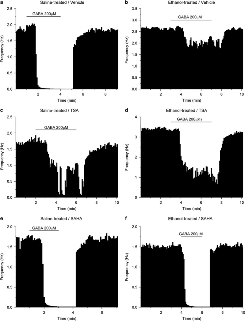
Figure 1.
Single neurons: effects of 200 μM GABA. Each figure represents the firing rate of a single neuron of the VTA over time. Vertical bars are proportional to the firing rate over a 5-s interval, horizontal bars represent the duration of application of GABA (200 μM). Neurons were recorded in brain slices from mice treated with saline (a, c, e) or ethanol (b, d, f). Slices were incubated with DMSO (a, b), TSA (c, d), or SAHA (e, f). Addition of 200 μM GABA to the extracellular medium resulted in maximum reductions of firing in these neurons of (a) −99.6%, (b) −22.7%, (c) −76.9%, (d) −70.0%, (e) −99.2%, (f) −98.5%.

