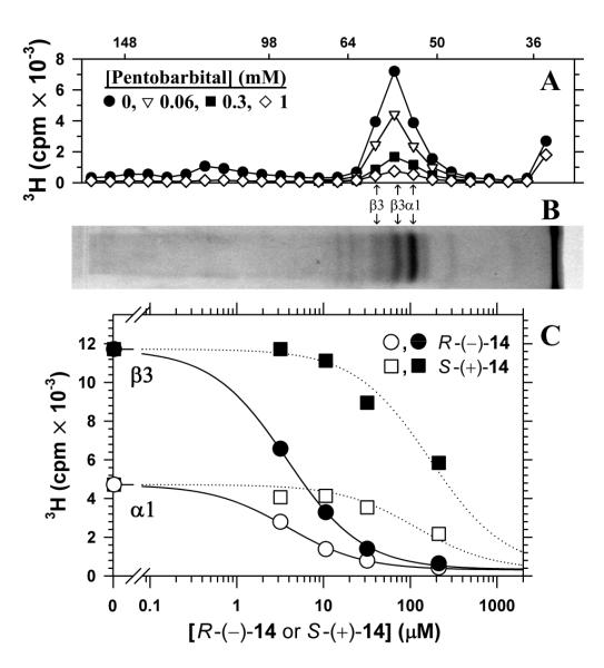
Figure 6.
Photolabeling of human α1β3 GABAAR with [3H]R-(−)-14 and concentration-dependent, stereospecific inhibition of photolabeling by non-radioactive barbiturates. Aliquots of purified human α1β3 GABAAR (~4 pmol [3H]muscimol sites in 180 μL final volume), equilibrated with 1 mM GABA, 0.3 μM [3H]R-(−)-14 (~2 μCi), and various concentrations of pentobarbital (A and B), R-(−)-14 (C), or S-(+)-14 (C), were irradiated (365 nm UV) for 30 min, and then GABAAR receptor subunits were isolated by SDS-PAGE as described in the Methods. A, 3H gel slice analysis (4 mm slices) of samples labeled in the presence of various concentrations of pentobarbital, with one of the Coomassie Blue-stained gel lanes shown in B. The migration of the molecular mass standards are indicated above the graph, and the known positions of the GABAAR α1 and β3 subunits21 are indicated below the graph with reference to the Coomassie Blue stained gel. For analysis of the concentration dependence of inhibition, the cpm in the single gel band containing the α1 subunit or the combined cpm in the two gel bands containing the β3 subunits were fit to a single site model (see Methods). Pentobarbital inhibited photolabeling of both the α1 and β3 subunits with IC50 = 88 ± 8, respectively, and background (bkg) values of 390 ± 110 cpm and 290 ± 255 cpm. C, Non-radioactive R-(−)-14 (●,○) inhibited [3H]R-(−)-14 incorporation into the α1 (○) and β3 (●) subunits with IC50 values of 3.8 ± 0.3 μM and 3.8 ± 0.2 μM, respectively, and bkg values of 310 ± 65 cpm and 330 ± 100 cpm. S-(+)-14 (■,□) inhibited α1 (□) and β3 (■) subunit photolabeling with IC50 values of 120 ± 30 μM and 170 ± 30 μM, respectively (dotted lines), with the values of bkg for each subunit fixed at those determined from the fit of the R-(−)-14 data.

