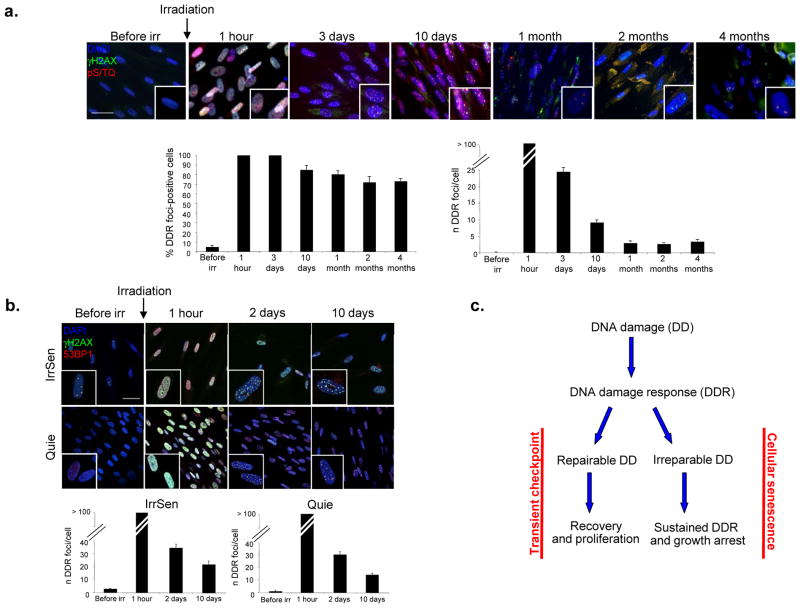
Figure 1. IR induces persistent DDR activation and cellular senescence.
a. IR generates persistent DDR. Top, images show DDR foci induction and resolution in early passage quiescent (contact-inhibited) BJ human fibroblasts following exposure to 20 Gy IR. Persistent DDR, in the form of γH2AX and pS/TQ foci, is still detectable even 4 months after IR. Bottom, bar graphs show the fraction of γH2AX foci-positive cells (± s.e.m.) (on the left) and the average number of γH2AX foci (± s.e.m.) per cell (on the right), at the indicated time points. More than 100 discrete foci cannot be counted accurately due to their proximity (1 hour time point). (For the quantification shown, around 100 cells per time point were analysed; scale bar = 20 μm) b. IrrSen cells are able to resolve additional IR-induced (10 Gy) DNA damage to an extent similar to Quie cells, as shown by the comparable kinetics of resolution of 53BP1 and γH2AX foci per cell over time after IR. (For the quantification shown, around 100 cells per time point were analysed; scale bar = 50 μm) c. Model, two opposite outcomes are possible upon DNA damage generation.

