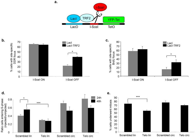
Figure 7. Ectopic TRF2 modulates DNA repair and DDR focus persistence, and exposed telomeric DNA ends cause a prolonged checkpoint.
a. Schematic of the integrated locus studied in NIH 2/4 cells37. Upon transfection, LacI or LacI-TRF2 binds to the lactose operator (LacO) repeats, YFP-Tet binds to the tetracycline operator (TetO) repeats, and RFP-I-SceI-GR cuts the specific site between the two sets of repeats. b. Quantification of cells positive for γH2AX at the I-SceI-locus (± s.e.m.) expressing LacI or LacI-TRF2, as detected by immunofluorescence confocal microscopy. I-SceI ON corresponds to 3 hours after RFP-I-SceI-GR induction, I-SceI OFF corresponds to 24 additional hours after removal of inducing agent. I-SceI site was detected as a distinct focus double-positive for YFP-Tet and anti-LacI antibody signals (* p value < 0.05; for the quantification shown, around 100 cells per sample were analyzed; n = 2). c. Quantification of cells positive for a BrdU signal (± s.e.m.) at the I-SceI-locus expressing LacI or LacI-TRF2, as detected by BrdU immunostaining under non-denaturing conditions and confocal microscopy. Values were normalized on the fraction of cells that had incorporated BrdU. (* p value < 0.05; for the quantification shown, around 100 cells per sample were analyzed; n = 2). d. Linearized telomeric DNA triggers a prolonged cell cycle arrest. Bar graph shows the ratio (± s.e.m.) between the percentages of injected cells that underwent DNA synthesis (assayed by BrdU incorporation) and the percentages of uninjected cells in the same experiment, at 24 or 48 hours after microinjection (* p-value < 0.05; *** p-value < 0.001; for the quantification shown, around 200–400 cells per time point, per DNA type were analyzed; n = 3). e. Linearized telomeric DNA impedes cell proliferation. Bar graph shows the percentages (± s.e.m.) of cells that underwent mitosis at 48 hours post-microinjection (*** p-value < 0.001; for the quantification shown, around 200 cells per DNA type were analyzed; n = 3). Passage through mitosis was monitored by detection in the cytoplasm of nucleus-injected IgG 48 hours after microinjection, as detected by immunofluorescence.

