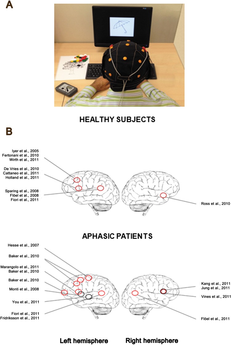
Figure 4.
(A) A patient with aphasia during an experimental session with transcranial direct current stimulation (tDCS): the anodal electrode is placed over the (left) perilesional area and the cathodal over the contralateral hemisphere. tDCS is delivered during speech therapy through a special stimulating cap that allows a simple positioning of electrodes. (B) Schematic diagrams of brain locations where tDCS improved language in normal subjects (top) and in aphasic patients (bottom). Red circles represent anodal stimulation and black circles cathodal stimulation (active electrodes). Note, however, that the position of the reference electrode differed across different studies. This figure is only reproduced in colour in the online version and red circles are grey in the printed version.

