
Figure 3.
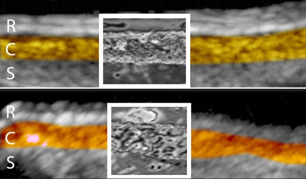
20 MHz ultrasound images of normal (top) and age-related macular degeneration (AMD) (bottom) eyes after semiautomated segmentation of retina (R), choroid (C) and sclera (S) as indicated by coloration. Wavelet processing was applied to the echo data encompassed by boxed regions. Note enhancement of druse in wavelet-processed area of AMD image. The choroid is more irregular in thickness in AMD than in normal eyes, as demonstrated here. These ‘hybrid’ or wavelet augmented ultrasound images demonstrate the use of classifiers such as interscale phase consistency.