FIG. 2.
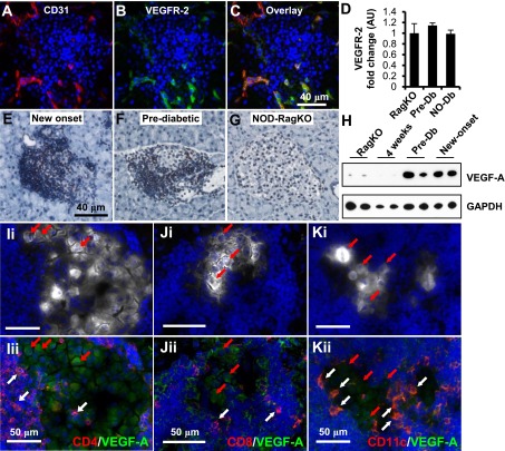
Insulitis promotes overexpression of VEGF-A. Immunofluorescence staining of CD31 (A, red), VEGFR-2 (B, green), and overlay (C). D: Quantification of VEGFR-2 transcripts from islet RNA at different stages of diabetes progression (n = 5 per group). NO-Db, new-onset diabetic; Pre-Db, prediabetic; RagKO, NOD RAG-deficient mice. Immunohistochemistry for VEGF-A performed on pancreatic sections from new-onset (E), prediabetic NOD (F), or NOD-RagKO female mice (G). H: Western analysis of whole-islet homogenates for VEGF-A from two mice per group. GAPDH, glyceraldehyde-3-phosphate dehydrogenase. I–K: Representative coimmunofluorescence staining of pancreata from diabetic NOD mice for insulin (white), VEGF-A (green), CD4 (I, red), CD8 (J, red), CD11c (K, red), and DAPI (blue). White arrows highlight examples of CD4+, CD8+, and CD11c+ cells that express VEGF-A. Red arrows indicate β-cells that express VEGF-A. Images i and ii for I–K are the same section with different signals depicted to highlight the islet area (white, Ii–Ki) and the VEGF-A+ cells (green, Iii–Jii).

