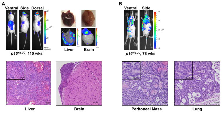
Figure 4. p16LUC Activity Detects Spontaneous Cancers In Vivo.

(A) (Top) Detection of a spontaneous histiocytic malignancy in a 110-week-old p16+/LUC mouse. All luminescent images shown were 1 min in length except the ventral image, which was 2 min long. Organ images were taken immediately following imaging. (Bottom) Haematoxylin and eosin stained fixed tissues confirm the presence of malignancy within luciferase positive regions of the brain and liver.
(B) (Top) Luminescent detection of a spontaneous, disseminated lung adenocarcinoma in a 78-week-old p16+/LUC mouse. Images were generated as in (A) with confirmatory histology shown below.
