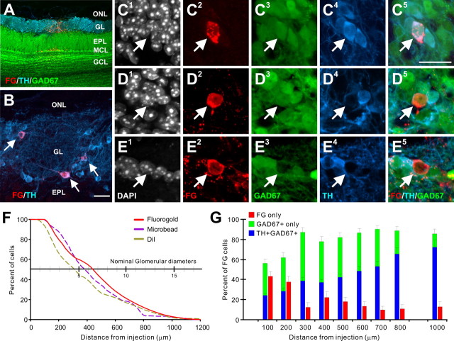
Figure 2.
Retrograde labeling of interglomerular neurons. A, Fluorogold-labeled cells (red) at a glomerular injection site in a GAD67-GFP (green) transgenic mouse, immunostained for TH (blue). A small number of mitral cells located below the injection site are labeled. B, FG-labeled (red) and TH-labeled (blue) juxtaglomerular neurons (arrows) 500 μm distant to a glomerular injection. C, FG+ cell within the glomerular layer 400–500 μm distant to the FG injection site expressing GAD67 and TH (C1 shows DAPI nuclear stain, C2 shows FG, C3 shows GAD67-GFP, C4 shows TH, and C5 shows triple overlay). D, FG+ cell within the glomerular layer 400–500 μm distant to the injection site expressing GAD67 but not TH. E, FG+ cell within the glomerular layer 400–500 μm distant to the injection site negative for GAD67 and TH. F, Percentage of FG-labeled cells plotted as a function of distance from the injection site. The distribution of labeled cells was not significantly different from previous DiI and microbead mouse glomerular injections (data from Aungst et al., 2003). G, Proportion of FG-labeled cells containing FG only, FG and TH/GAD67, or FG and GAD67 as a function of distance from the injection site. ONL, Olfactory nerve layer; MCL, mitral cell layer; GCL, granule cell layer. Scale bars: A (in B), 20 μm; B, 10 μm; C–E (in C), 20 μm.

