Figure 4.
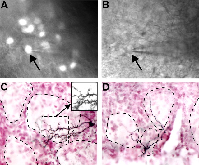
Biocytin filling of TH+GAD67+ cells in the glomerular layer. A, Neurons expressing GFP are easily visualized in the glomerular layer using an epifluorescent microscope. B, The same cell is readily visible in DIC optics allowing whole-cell patch-clamp recording and biocytin filling of identified neurons (patch pipette attached to GFP+ cell, arrow in A and B). C, D, Biocytin-filled cell processes from two different cells stained with NiDAB and glomerular cellular boundaries visualized with neutral red staining. Inset, Biocytin-filled cell dendrites in the absence of neutral red staining. Dotted lines indicate outline of glomerular neuropil.

