Fig. 1.
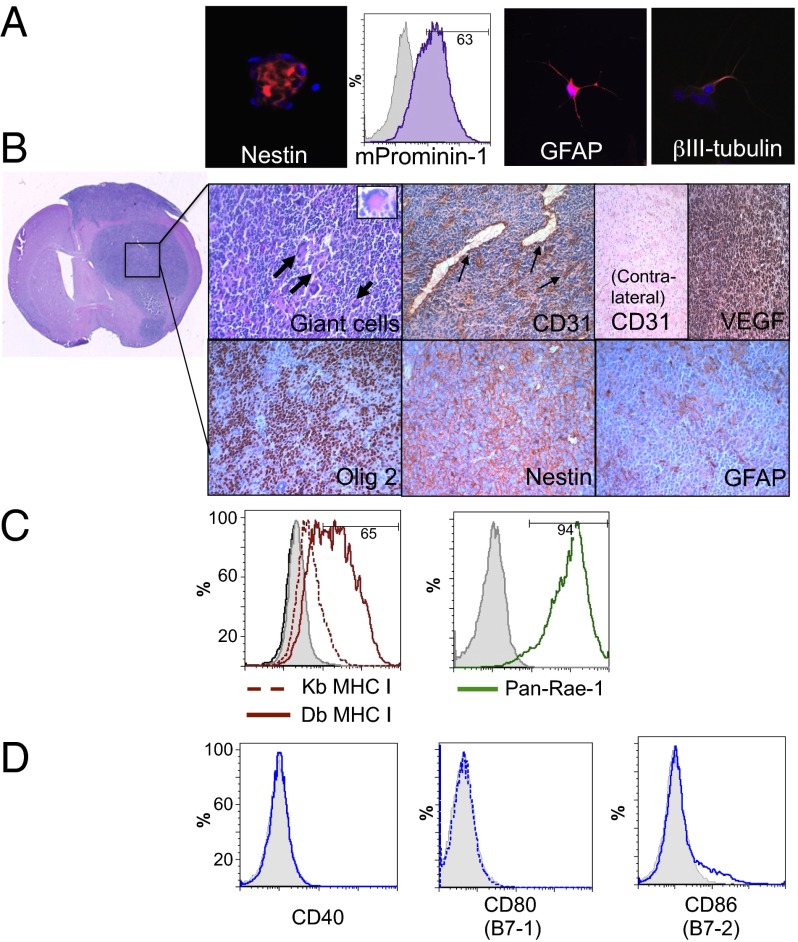
In vitro and in vivo characterization of mouse 005 GSCs. (A) Neurospheres express the stem cell markers nestin (red; Left) and mProminin-1 (CD133 homolog; FACS, Center Left) and can be differentiated in serum-containing media into glial (GFAP; red, Center Right) and neuronal (βIII-tubulin; red, Right) phenotypes. (B) Murine 005 GSCs form characteristic gliomas in C57BL/6 mice. Pleomorphic, multinucleated giant cells seen by H&E staining (bold arrows and Inset; Upper Left) along with dense CD31-positive dilated vasculature (arrows) are observed within the tumors and not on the contralateral side of the brain. Murine 005 tumors also express VEGF and maintain stem cell characteristics, as seen by immunohistochemical staining (brown) of Olig2 and Nestin, and limited heterogeneous expression of GFAP (Lower Right). (C) In vitro, 005 GSCs only express MHC class I after treatment with IFN-γ (solid-Db and dotted-Kb red line), but strongly express NK ligand Rae-1 (Pan-Rae; solid green line). (D) Murine 005 GSCs lack expression of costimulatory signaling molecules CD40 and CD80 and contain only a minor population of CD86 (blue lines). Isotype controls, gray filled.

