Fig. 3.
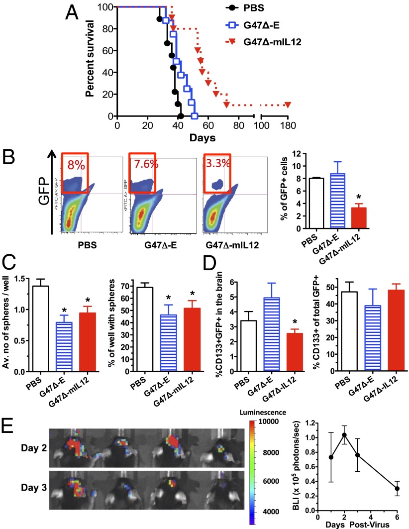
Treatment of established intracranial 005 tumors in C57BL/6 mice. (A) Murine 005 GSCs (5 × 10⁴ cells) were stereotactically implanted and mice treated with intratumoral injections of PBS, G47Δ-E, or G47Δ-mIL12 (n = 10) on days 8 and 12. Kaplan–Meier analysis shows a significant extension in survival with G47Δ-mIL12 treatment, with a median survival of 57 d compared with G47Δ-E (41 d) or PBS (37 d). (B) Representative flow cytometry results of GFP+ 005 cells (quadrant bordered in red; Left) extracted from brains on day 15. A significant decrease in tumor burden was observed after G47Δ-mIL12 treatment (*P < 0.05; Right graph). (C) GFP+ 005 GSCs were sorted from brain tumors and plated at 10–30 cells per well for self-renewal assays. Both G47Δ-E and G47Δ-mIL12 similarly (P = 0.3) decreased the ability of GSCs to form spheres, number per well, and percent sphere-positive wells versus PBS (*P < 0.05). (D) There is no difference when comparing treated and PBS in the percentage of CD133+ cells within the GFP+ population in tumors on day 15. The percent of CD133+ GFP+ cells after G47∆-mIL12 is significantly less than G47∆-E (*P < 0.05). (E) Bioluminescent imaging of virus replication in vivo. Established 005 tumors were injected with G47∆-Us11fluc on day 13 postimplantation of 105 cells. Left, images of four mice in the prone position on days 2 and 3 postinfection, with heat scale on Right (radiance; photons per s/cm2 per steradian). Right, mean luminescence (supine position) from four mice. Error bars indicate SEM.

