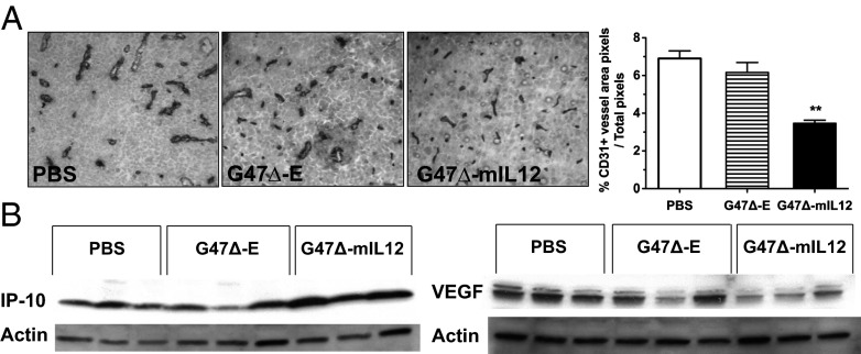
Fig. 5.
Antiangiogenic effects of G47Δ-mIL12 in vivo. (A) Immunohistochemical staining of endothelial cells (anti-CD31 antibody) on brain tumor sections 6 d after treatment with PBS, G47Δ-E, and G47Δ-mIL12 (day 18). Right, histogram of CD31+ vessel area, as determined by ImageJ analysis (n = 3 per group). Only G47Δ-mIL12 treatment significantly decreased CD31+ microvessel density (**P < 0.005 versus G47Δ-E). Error bars indicated SEM. (B) Western blot of lysates from treated brain tumors on day 18 demonstrated an increase in IP-10 (Left) and a decrease in VEGF expression (Right) after G47Δ-mIL12 treatment. Each lane represents an individual mouse.

