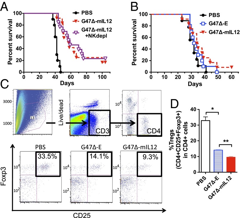
Fig. 6.
Role of T cells in G47Δ-mIL12 treatment of intracranial 005 GSC tumors. (A) NK-cell depletion had no effect on the ability of G47Δ-mIL12 to enhance survival (P < 0.001; G47Δ-mIL12 with (G47∆-mIL12 + NKdepl) and without NK depletion (G47∆-mIL12) compared with PBS; P = 0.7, G47Δ-mIL12 with NK depletion versus without). (B) 005 GSCs were implanted in athymic mice, devoid of T cells, and treated with G47Δ-E or G47Δ-mIL12 on days 8 and 12. No significant difference was observed between these two treatments or compared with PBS. (C) FACS analysis of cells from treated 005 GSC tumors in C57BL/6 mice on day 6 posttreatment. Gates were drawn on live CD3+ cells, followed by CD4+ (Upper), and then the double-positive CD25+Foxp3+ cells were defined as Tregs. Representative flow cytometry plots of Tregs (black boxes, Lower) after PBS, G47Δ-E, and G47Δ-mIL12 treatment. (D) Significant decrease in Tregs (percent of CD4+) was observed with G47Δ-E versus PBS (**P < 0.005), and a further significant reduction with G47Δ-mIL12 versus G47Δ-E (**P < 0.005). Error bars indicate SEM.

