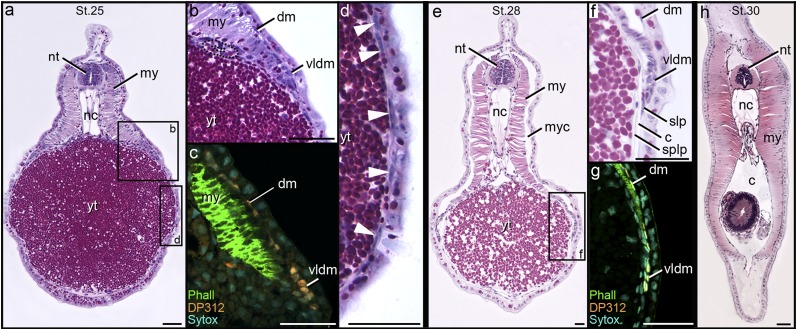
Fig. 2.
Plastic sections and DP312 labeling of Petromyzon embryos and larvae. (A, B, D–F, and H) Plastic sections stained with H&E. (C and G) Cryosections are labeled for Pax3/7 (orange; DP312), skeletal muscle (green; phalloidin), and nuclei (cyan; Sytox). (A–D) Stage 25. DP312-positive cells of the DM (dm) are adjacent to the inner surface of the ectoderm and form an epithelial lip ventrally (vldm; compare B with C). Elongate cells of the presumptive LPM (arrowheads in D) surround the yolk tube (yt). (E–G) Stage 28. The LPM has split into somatic (slp) and splanchnic (splp) layers, forming a coelom (c). (E–G) The ventral lip of the dermomyotome is positioned between ectoderm and somatic lateral plate. (H) Stage 30. Myotomes close the body wall ventrally. Fig. S2 shows approximate planes of section. my, myotome; myc, myocoel; nc, notochord; nt, neural tube. (Scale bar: 50 µm.)

