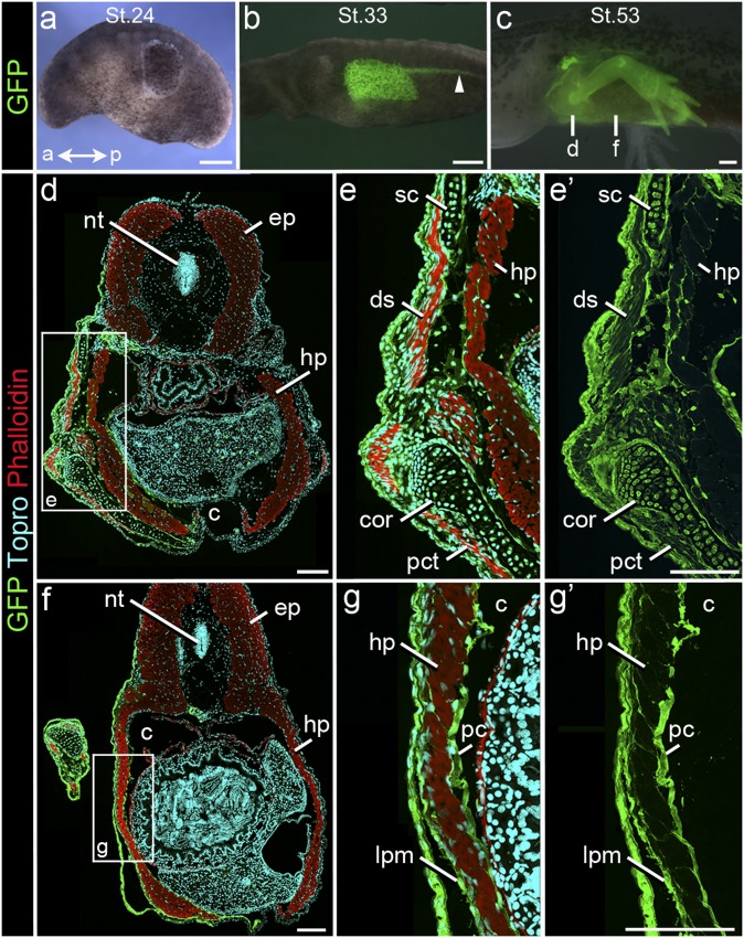
Fig. 5.
GFP to WT Isotopic transplants of LPM/Pronephros/Ect in Ambystoma. (A–C) Lateral view of a surgery chimera at (A) stage 24 (time of surgery) and the same specimen at (B) stages 33 and (C) 53, showing distribution of GFP donor tissue (green). Note (B) caudal migration of pronephros (arrowhead) and (C) graft-derived skeletal elements of the forelimb. (Scale bar: 500 µm.) (D–G′) Cryosections through (D–E′) the pectoral and (F–G′) the interlimb regions of larva pictured in C labeled for GFP (green), skeletal muscle (red; phalloidin), and nuclei (cyan; TO-PRO). (D–E′) In the pectoral region, graft-derived cells are present in scapula (sc) and coracoid (cor) and form the connective tissue of appendicular muscles (ds, dorsalis scapulae; pct, pectoralis) and the hypaxial myotome (hp). (F–G′) In interlimb levels, graft-derived cells form mesenchyme lateral to hp as well as connective tissue within hp. ep, epaxial myotome; pc, parietal coelomic lining. Fig. 2 defines abbreviations. (Scale bar: 250 µm.)

