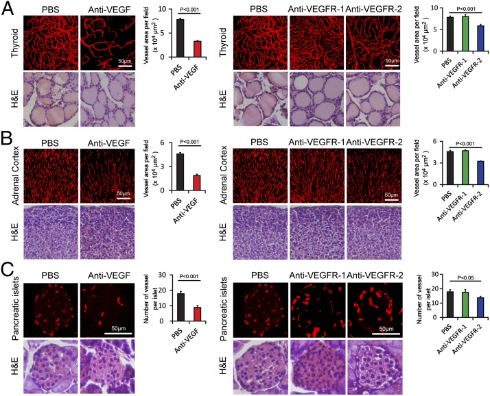
Fig. 1.
Impact of anti-VEGF blockades on vasculature in endocrine organs. (A) CD31+ thyroid microvessels (red). H&E staining was used to reveal tissue structures. Vessel areas were quantified (20× magnification, n = 8 fields per group). (B) Endomucin+ adrenal cortex microvessels were detected in paraffin-embedded tissues (red). H&E staining was used to reveal tissue structures. Vessel areas were quantified (20× magnification, n = 8 fields per group). (C) Endomucin+ pancreatic islet microvessels were detected in paraffin-embedded tissues (red). H&E staining was used to reveal tissue structures. Vessel numbers were quantified (20× magnification, n = 8 fields per group). Data are presented as means ± SEM.

