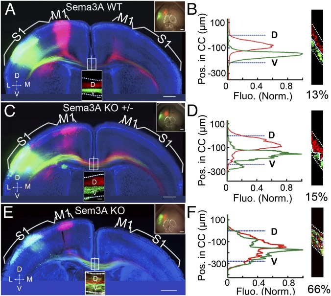
Fig. 4.
Disruption of axon order in Sema3A-KO mice was specific and dosage dependent. (A and B) A 200-µm coronal section from a P8 WT mouse brain (littermate of the Sema3A KO mouse) with mCherry expression in M1 and EGFP expression in S1, presented as in Fig. 1 A and B. As shown by the mean fluorescence intensity of M1 and S1 axons along the D–V axis of the CC and the percentage of overlap at the midline, the axon order was not disrupted. (C and D) A 200-µm coronal section of the brain of a P8 Sema3A-heterozygous mouse (from the same litter as the Sema3A KO mice) with mCherry expression in M1 and EGFP expression in S1. No disruption of axon order in the CC was observed. (E and F) A 200-µm coronal section from the brain of a P8 Sema3A KO mouse with mCherry expression in M1, and EGFP expression in S1. The D–V order within the CC was grossly disrupted in the Sema3A KO mice, as shown by the mean fluorescence intensity of M1 and S1 axons along the D–V axis at the midline and the high percentage of overlap shown in F. (Scale bars, 500 μm in A, C, and E.) Dotted white lines indicate the CC border.

