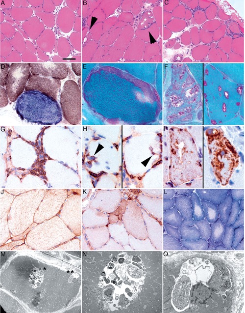
Figure 1.

Muscle biopsy from the right lateral vastus muscle showing atrophic fibers with caliber changes (A), many rimmed vacuoles together with endomysial inflammatory infiltrates (B, C), COX negative-fibers (D) and a single RRF with rimmed-vacuoles (E, F). Inflammatory infiltrates consisted mainly of CD3-positive T-cells, including CD8-positive cytotoxic T-cells with invasion of intact muscle fibers (G, H). SMI-31 (neurofilament) and p62 (I) showed immunoreactivity in the vacuoles. Signs of a neurogenic muscle lesion with fiber-type grouping and upregulation of NCAM were noted (K). Ultrastructural examination revealed fibers with autophagic vacuoles with sequestered cytoplasmic organelles and degradation products (M, N, O).
