
Figure 3.
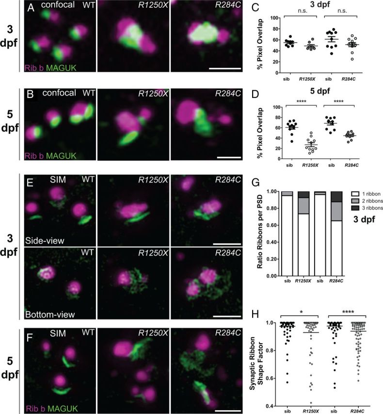
cav1.3a mutant hair-cell synapses progressively lose juxtaposition of presynaptic and postsynaptic components. A, B, Representative confocal images of Ribeye b (Rib b) and MAGUK in 3 dpf (A) and 5 dpf (B) WT and cav1.3a mutant hair cells. For display purposes, images were resampled (bicubic) in Photoshop to minimize pixilation. Scale bars, 1 μm. C, D, Percentage of MAGUK-label containing pixels overlapping with Ribeye b in 3 dpf (C) and 5 dpf (D) WT and cav1.3a mutant NM hair cells. Each circle represents an NM in an individual larva. Error bars are SEM. C, MAGUK immunolabel overlapped with Ribeye b comparably in 3 dpf mutants and WT. Unpaired t test, p = 0.1811 and 0.1162, respectively. D, MAGUK immunolabel overlapped significantly less with Ribeye b in 5 dpf mutants. Unpaired t test, ***p < 0.0001 for both mutant alleles. E, F, SR-SIM images of ribbon synapses in 3 dpf (E) and 5 dpf (F) cav1.3a mutants and WT. Scale bars, 1 μm. E, Images of 3 dpf ribbon synapses. The synaptic ribbons in cav1.3a mutants appear enlarged and often misshapen. The bottom-view images show MAGUK label beneath the ribbon synapse. F, Images of 5 dpf ribbon synapses. MAGUK appears even less spatially restricted to the synaptic ribbon than in 3 dpf hair cells. G, Fraction of 3 dpf ribbon synapses within individual NMs with PSDs juxtaposing one, two, or three synaptic ribbons. cav1.3a mutant NMs synapses contain two to three synaptic ribbons with much greater frequency than WT (n = 4 NMs per condition, each containing ∼15–25 synapses). H, The shape factor of synaptic ribbons in 3 dpf hair cells. Each spot represents an individual ribbon. The horizontal bars represent the mean values. Synaptic ribbons are significantly less round in cav1.3a mutants than WT siblings (sib). Mann–Whitney U test, *p = 0.0266 and ****p < 0.0001, respectively.